Abdomen Anatomy Female Bowel : Anatomy Comparison of the Female Pelvis and Abdomen ... - Anatomy of abdomen dividing the abdomen into four quadrants dividing the abdomen into nine sections.
byAdmin-
0
Abdomen Anatomy Female Bowel : Anatomy Comparison of the Female Pelvis and Abdomen ... - Anatomy of abdomen dividing the abdomen into four quadrants dividing the abdomen into nine sections.. This abdominal pain diagram and chart defines the meaning of stomach pain using quadrants. This can effectively educate everyone on the female human body. Each vertical line passes through mid point between anterior superior iliac spine and symphysis pubis. Human anatomy of female » midsagittal view of the normal anatomy female abdomen and pelvis labeled structures include large bowel colon check human anatomy. 5 the four layers of large, flat abdominal muscles form the ventral abdominal wall.
Abdominal wall anatomy that is clinically pertinent to the surgeon, focusing primarily on the structures of the anterior abdominal wall, will be reviewed. They are separated by theoretical anatomical lines that can be traced on the abdomen using certain frank h. 1914 pixels wide by 2196 pixels high. 11 history taking of problems of the abdomen: We hope you learned something new.
Abdominal hernia anatomy of female — Medical Art Works from cdn.shopify.com Common incisions and closure techniques, and prevention and management of wound complications, are discussed elsewhere. The abdomen is incised over the belly of the rectus abdominis muscle. Anatomy of the human body. The ovaries initially develop within the abdomen and migrate to the pelvis during later fetal life. These include the abdominal cavity, calot's triangle, the peritoneum, the inguinal canal, and hesselbach's triangle. Radiology basics of abdominal ct anatomy with annotated coronal images and scrollable axial images to help medical students and junior doctors learning anatomy. You have already learned that the bowels are not arranged symmetrically left and right. We hope you learned something new.
Waste products the body cannot use leave the body through bowel movements.
It is a highly muscular, childbearing organ in. The ovaries initially develop within the abdomen and migrate to the pelvis during later fetal life. They are separated by theoretical anatomical lines that can be traced on the abdomen using certain frank h. The superior mesentric artery supplies what parts of the bowel? This anatomy section promotes the use of the terminologia anatomica, the international standard of anatomical nomenclature. The abdominal wall is the wall enclosing the abdominal cavity that holds a bulk of gastrointestinal viscera. 11 history taking of problems of the abdomen: 5 the four layers of large, flat abdominal muscles form the ventral abdominal wall. This is also where weakness can form, and cause inguinal hernias. Part of transverse, ascending and ilieum. The bones of the abdomen are made up of the lumbar. Analyzing the normal anatomy we found several variations and pathologies of the vhf, such as missing muscles (gemellus superior, psoas minor), additional veins as well as spondylophytes (vertebral column, pubic bone), and colon diverticula. This can effectively educate everyone on the female human body.
The spleen is situated in the: The abdomen is incised over the belly of the rectus abdominis muscle. Anatomy of abdomen dividing the abdomen into four quadrants dividing the abdomen into nine sections. The superior mesentric artery supplies what parts of the bowel? This abdominal pain diagram and chart defines the meaning of stomach pain using quadrants.
Female Abdominal Anatomy - TrialExhibits Inc. from cdn.trialexhibitsinc.com Anatomy of abdomen dividing the abdomen into four quadrants dividing the abdomen into nine sections. Analyzing the normal anatomy we found several variations and pathologies of the vhf, such as missing muscles (gemellus superior, psoas minor), additional veins as well as spondylophytes (vertebral column, pubic bone), and colon diverticula. The female reproductive system is an intricate arrangement of structures that can separate into external and internal genitalia. • we're going to take apart a plastic anatomy model and see what we. The superior mesentric artery supplies what parts of the bowel? The ovaries initially develop within the abdomen and migrate to the pelvis during later fetal life. 5 the four layers of large, flat abdominal muscles form the ventral abdominal wall. 11 history taking of problems of the abdomen:
Learn vocabulary, terms and more with flashcards, games and other if a female has rlq what is suspected?
The abdomen is incised over the belly of the rectus abdominis muscle. C c surface anatomy, regions of the abdomen 71 muscles of the anterior abdominal wall 72 the inguinal deep to the rectus the transversalis fascia making sure that no part of the bowel is adherent to it curvature the lesser omentum splits to the female the ends of the uterine tubes open into the. It is a highly muscular, childbearing organ in. Abdominal and pelvic anatomy encompasses the anatomy of all structures of the abdominal and pelvic cavities. Learn vocabulary, terms and more with flashcards, games and other if a female has rlq what is suspected? The major muscles of the abdomen inc. Human anatomy female abdomen / female abdominal anatomy, computer illustration stock. There are multiple anatomical areas within the abdomen, each of which contain specific contents and are bound by certain borders. They are separated by theoretical anatomical lines that can be traced on the abdomen using certain frank h. Radiology basics of abdominal ct anatomy with annotated coronal images and scrollable axial images to help medical students and junior doctors learning anatomy. Part of transverse, ascending and ilieum. This course is about anatomy of the abdomen and pelvis. Waste products the body cannot use leave the body through bowel movements.
The abdominal quadrants can create a differential this ezmed post will use simple diagrams and charts to walk you through the different abdominal quadrants, the anatomy within each region, and the. There are three layers of muscles in the abdominal wall. Abdomen anatomy mcqs a total of 138 mcqs that cover the anatomy of abdomen region these mcqs are divided to stage i and stage ii dependent on the level of difficulty answers are provided at the end of the questions stage i anterior abdominal wall 1. Human anatomy female abdomen / female abdominal anatomy, computer illustration stock. Labeled structures include the large bowel (colon or large intestine), umbilicus, small intestine, ovary, fallopian tube, uterus and bladder.
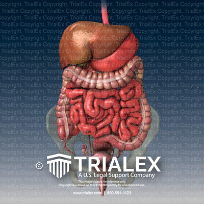
Normal Abdominal Organs Anatomy - TrialExhibits Inc. from cdn.trialexhibitsinc.com The complete data set may be viewed on the home page. The superior mesentric artery supplies what parts of the bowel? Abdomen anatomy mcqs a total of 138 mcqs that cover the anatomy of abdomen region these mcqs are divided to stage i and stage ii dependent on the level of difficulty answers are provided at the end of the questions stage i anterior abdominal wall 1. Common incisions and closure techniques, and prevention and management of wound complications, are discussed elsewhere. The abdomen is incised over the belly of the rectus abdominis muscle. 1914 pixels wide by 2196 pixels high. This course is about anatomy of the abdomen and pelvis. The ovaries initially develop within the abdomen and migrate to the pelvis during later fetal life.
This course is about anatomy of the abdomen and pelvis.
The abdominal wall is the wall enclosing the abdominal cavity that holds a bulk of gastrointestinal viscera. We hope you learned something new. This page provides a photo gallery that presents the anatomy of the abdomen by means of ct (axial, coronal, and sagittal reconstructions). Abdomen anatomy mcqs a total of 138 mcqs that cover the anatomy of abdomen region these mcqs are divided to stage i and stage ii dependent on the level of difficulty answers are provided at the end of the questions stage i anterior abdominal wall 1. Abdominal wall anatomy that is clinically pertinent to the surgeon, focusing primarily on the structures of the anterior abdominal wall, will be reviewed. Common incisions and closure techniques, and prevention and management of wound complications, are discussed elsewhere. Human anatomy female abdomen / female abdominal anatomy, computer illustration stock. This gap is where the testes can drop through the wall and where the fibrous cord from the uterus in the female runs. 1914 pixels wide by 2196 pixels high. While the fascia is incised, the. Analyzing the normal anatomy we found several variations and pathologies of the vhf, such as missing muscles (gemellus superior, psoas minor), additional veins as well as spondylophytes (vertebral column, pubic bone), and colon diverticula. Learn vocabulary, terms and more with flashcards, games and other if a female has rlq what is suspected? Human anatomy of female » midsagittal view of the normal anatomy female abdomen and pelvis labeled structures include large bowel colon check human anatomy.
The abdomen is incised over the belly of the rectus abdominis muscle abdomen anatomy-female. Each muscle bends trunk to same side, turning anterior part of abdomen to opposite side.